LipAge Protocol:
viability after thawing
The question physicians and patients ask
When a physician introduces LipAge, a question often comes up: “Will the sample still be in good condition after years of frozen storage? Do the cells survive thawing?”
It is a legitimate question. What matters is not just cell survival — it is the maintenance of biological function: differentiation capacity, extracellular vesicle integrity, and preservation of the secretory profile. This document presents photomicrographic evidence produced in the Cryopraxis laboratory.
The images in this edition come from actual adipose tissue samples processed and cryopreserved using the LipAge Protocol, then thawed and assessed in the laboratory. They show: (1) osteogenic differentiation of mesenchymal cells from thawed adipose tissue and (2) morphological integrity of extracellular vesicles after thawing. The photomicrographs are the intellectual property of Cryopraxis®.
Photomicrographic evidence — Cryopraxis laboratory
The two images below were obtained from samples that underwent the complete LipAge Protocol: collection, processing, cryopreservation in liquid nitrogen, and controlled thawing.
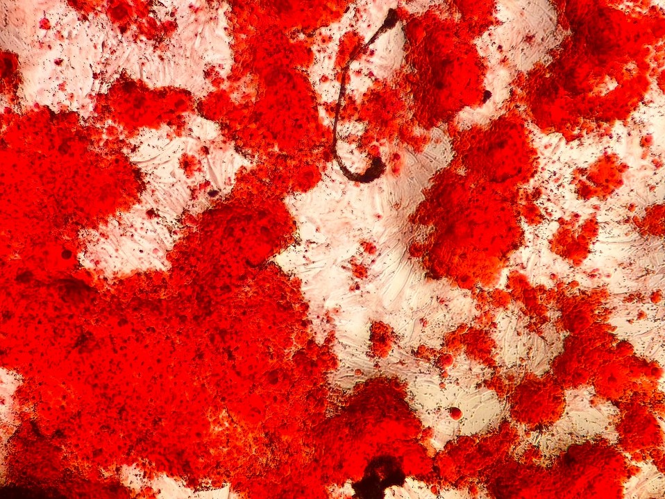
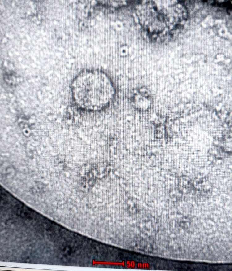
Figure 1 — Osteogenic differentiation (Alizarin Red S)
Alizarin Red S staining highlights calcium deposits (mineralization nodules) in red-orange — a marker of successful osteogenic differentiation. The abundance of nodules indicates that mesenchymal cells from cryopreserved adipose tissue retained their capacity for osteogenic differentiation after thawing.
Relevance: osteogenic differentiation is one of the three classic capabilities of these cells (alongside adipogenic and chondrogenic differentiation) and a robust indicator of preserved function.
[FACT] Alizarin Red S-positive nodules are an internationally accepted histochemical criterion for confirming osteogenic differentiation (Dominici et al., 2006; Pittenger et al., 1999).
Figure 2 — Extracellular vesicles imaged by TEM
Transmission electron microscopy is the reference method for the morphological characterization of extracellular vesicles. The image shows structures with intact lipid bilayer membranes, spherical morphology, and diameters between 30 and 100 nm, consistent with the criteria of the International Society for Extracellular Vesicles (ISEV).
Relevance: membrane integrity is a prerequisite for vesicles to transport miRNAs, growth factors (VEGF, HGF, TGF-β), and bioactive proteins.
[FACT] TEM characterization is a MISEV2018 requirement for the morphological confirmation of extracellular vesicles (Théry et al., 2018).
Why cryopreservation preserves function
Post-thaw viability is the result of a protocol developed and implemented at Cryopraxis's Cell Technology Center licensed by ANVISA (Brazil's health regulatory agency). Each step aims to minimize cellular damage from the thermal cycle:
- Controlled collection: the LipAge kit keeps the sample in a sterile environment at an appropriate temperature until it reaches the laboratory.
- Rapid processing and fractionation: reduce the time between collection and freezing.
- Validated cryopreservation: cryoprotectants at controlled concentrations and a controlled freezing curve (-1 °C/min), which prevents intracellular crystal formation.
- Storage in liquid nitrogen (-196 °C): drastically reduces metabolic activity, maintaining the material in its condition at the time of collection.
- Controlled thawing: rapid warming (37 °C) to minimize recrystallization.
- Post-thaw quality control (LipAge Score): morphology, viability (Trypan Blue), and microbiological safety before release.
Unlike adipose tissue in the body, material stored in liquid nitrogen remains biologically stable. Studies of materials cryopreserved for more than 20 years report recovery rates comparable to those seen with short-term storage, provided that processing and storage are rigorous.
Summary of evidence by fraction
| Whole adipose tissue (LipAge Fill) | Alizarin Red S / Oil Red O / H&E — adipocytes and precursors with preserved morphology; osteogenic and adipogenic differentiation confirmed after thawing. |
| Stromal fraction | Flow cytometry (CD34+/CD73+/CD90+/CD105+) — preserved mesenchymal profile; viability above 70% by Trypan Blue exclusion. |
| Extracellular vesicles | TEM (50 nm scale) + NTA — intact lipid bilayer membrane; diameter of 30–100 nm. Research area, with no clinical use. |
| Mesenchymal cells | MTT / Trypan Blue / cytometry — preserved viability; secretory capacity (VEGF, HGF) maintained in short-term studies. Research area, with no clinical use. |
What physicians need to know in practice
Every sample comes with a LipAge Score
Before any release, the sample undergoes quality control and receives a grade (A+ to D). The physician receives a report detailing the parameters assessed. A sample with microbiological contamination is not released, regardless of the other results.
Viability above 70% as an operational threshold
The minimum viability threshold for release under the LipAge Protocol is 70% viable cells by Trypan Blue exclusion, in line with ISCT and AABB standards. Samples below this threshold receive a C or D grade and follow the nonconformity protocol.
Extracellular vesicles: assessment under development
Full characterization of extracellular vesicles (NTA + TEM + proteomics) is not yet part of the routine LipAge Score assessment. The images in this edition are the result of internal Cryopraxis research.
Delivery of the material
Following controlled thawing and approval based on the LipAge Score, the material is delivered to the attending physician with chain-of-custody, traceability, and quality control documentation.
Bibliographic References
- Théry C et al. Minimal information for studies of extracellular vesicles 2018 (MISEV2018). J Extracell Vesicles. 2018;7(1):1535750.
- Dominici M et al. Minimal criteria for defining multipotent mesenchymal stromal cells. Cytotherapy. 2006;8(4):315-317.
- Pittenger MF et al. Multilineage potential of adult human mesenchymal stem cells. Science. 1999;284(5411):143-147.
- Baglioni S et al. Adipose tissue-derived progenitor cells and chronic wound healing. Front Med. 2024;11:1417920.
- Wang Z et al. Regulatory role of adipose tissue-derived exosomes in metabolic homeostasis. Front Endocrinol. 2024;15:1374715.
- Guo J et al. Long-term cryopreservation of adipose-derived stem cells: viability, proliferation and multilineage differentiation. Cryobiology. 2018;80:164-169.
- Zimmerlin L et al. Stromal vascular progenitors in adult human adipose tissue. Cytometry A. 2010;77(1):22-30.
- Patel DM et al. Characterization of human adipose-derived stem cells after cryopreservation and re-plating. Cytotherapy. 2013;15(11):1383-1391.
- AABB Standards for Cellular Therapy Services. 9th Edition. 2022.
- ANVISA. RDC 508/2021 — Boas práticas para o processamento de tecidos, células e órgãos humanos.